
 Medial Side
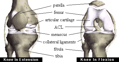
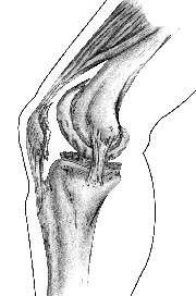
Ligaments help to stabilize the knee. The collateral ligaments run along the sides of the knee and limit sideways motion. The anterior cruciate ligament, or ACL, connects the tibia to the femur at the center of the knee. Its function is to limit rotation and forward motion of the tibia. (A damaged ACL is replaced in a procedure known as an ACL Reconstruction.) The posterior cruciate ligament, or PCL (located just behind the ACL) limits backward motion of the tibia.
These components of your knee, along with the muscles of your leg, work together to manage the stress your knee receives as you walk, run and jump.
Dr. Herbert D. Huddleston Presents: Arthritis of the Knee Joint
| ANATOMY OF THE NORMAL KNEE JOINT |
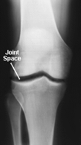
The knee is a "hinge type" joint which is formed by two bones held together by flexible ligaments. The bones are the femur (thigh bone) and the tibia (shin bone). The knee cap (patella) also forms part of the knee joint. It glides over the end of the femur as the knee bends. The moving parts of a normal knee are covered with a layer of articular cartilage which is a white smooth substance about 1/4 of an inch thick on the patella and 1/8 of an inch thick on the femur and tibia. An x-ray of the knee normally shows space (the "joint space") between the femur and the tibia as well as between the femur and the patella. This is not empty space but represents the cartilage (which does not show up on x-rays). The smooth, cartilage-covered surfaces of the knee move on each other with very little friction in the normal joint. In the normal knee the "joint space" is approximately 1/4 of an inch wide and fairly even in outline.
  |
Dr. Herbert D. Huddleston Presents: Arthritis of the Knee Joint
| DISEASES OF THE KNEE JOINT |
There are a number of conditions which can cause arthritis of the knee. The term arthritis literally means inflammation of a joint, but is generally used to describe any condition in which there is damage to the cartilage. Inflammation, if present, is in the synovium. The proportion of cartilage damage and synovial inflammation varies with the type and stage of arthritis. Usually the pain early on is due to inflammation. In the later stages, when the cartilage is worn away, most of the pain comes from the mechanical friction of raw bones rubbing on each other.
  |
| There are two broad categories of arthritis: OSTEOARTHRITIS AND RHEUMATOID ARTHRITIS |
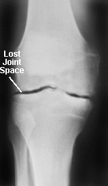
Osteoarthritis mainly damages the joint cartilage, but there is often some inflammation as well. It usually affects only one or two major joints (usually in the legs). It does not affect the internal organs. The cause of knee osteoarthritis is not known. It is thought to be simply a process of мwear and tearо in most cases. Some conditions may predispose the knee to osteoarthritis, for example, a previous fracture that involved the joint, or by lesser injuries that may have torn ligaments or menisci. Abnormalities in development of the knee bones, such as bow legs, may cause the knee to wear out sooner than normal. In osteoarthritis of the knee the cartilage cushion is either thinner than normal (leaving bare spots on the bone), or completely absent. Bare bones grind against each other and cause mechanical pain. Fragments of cartilage floating in the joint may cause inflammation in the joint lining, and this is a second source of pain. X-rays show the joint space to be narrowed and irregular in outline. There is no blood test for osteoarthritis.Rheumatoid Arthritis (R.A.) starts in the synovium and is mainly inflammatory. The cause is not known. It eventually destroys the joint cartilage. Bone next to the cartilage is also damaged, making it very soft. R.A. affects multiple joints simultaneously. It also affects internal organs. Another form of knee arthritis that is mainly мinflammatoryо is Lupus. There are other more rare forms of arthritis that are also mainly мinflammatoryо. They are basically similar to R.A.. X-ray changes in R.A. are essentially similar to osteoarthritis plus a loss of bone density.
Blood tests for rheumatoid arthritis are not very accurate. Rheumatoid Factor is present in the blood in about 80% of patients who have had rheumatoid arthritis for more than 18 months. Early on in the disease the percentage is much lower. Unfortunately, about 7% of people over the age of 70 test positive for rheumatoid factor, even though they do not have rheumatoid arthritis. The test, by itself, is therefore not very reliable.
Anti-inflammatory medications are effective in treating the inflammatory aspect of either rheumatoid or osteoarthritis.
Osteonecrosis is another (rare) condition which may cause knee pain. It is a condition in which parts of the femur bone die and later collapse.
| MENISCAL INJURIES |
Many patients have knee pain coming from injury to a meniscal cartilage rather than injury to the articular cartilage. Most people are not aware that there are these two types of cartilage in the knee. This is somewhat confusing. The articular cartilage is the cartilage that covers the ends of the bone (similar to the tread on a tire). A meniscal cartilage is a disc of cartilage that is actually separate from the femur and the tibia and the patella. There are two such c-shaped meniscal cartilages in the knee. They are sandwiched between the femur and the tibia. These meniscal cartilages are often injured, particularly during athletics.
If a meniscal cartilage is torn, it often does not heal and the pieces of the cartilage may become trapped in abnormal positions in the knee causing giving way, fluid on the knee, and pain with certain twisting activities. The arthroscope, which is an instrument the size of a pencil, can be inserted into the knee through a minute incision allowing the physician to visualize the contents of the knee on a television screen. With small instruments placed into the knee through other minute puncture wounds, the surgeon can often remove the torn bits of meniscal cartilage and relieve the problems described above ("Arthroscopic Surgery"). However, when the articular cartilage has been worn out (as in arthritis), arthroscopy is rarely able to correct the problem and a knee replacement is often needed.
http://www.fauxpress.com/kimball/med/ortho/kneeanat.htm
Комментариев нет:
Отправить комментарий